
TRANSLATIONAL RESEARCH
Ask more biomarker questions than limited tissue can answer.
Translational programs increasingly need to distinguish small differences in biomarker expression, not just detect presence or absence, and conventional IHC wasn't built for that resolution.
Virtual staining resolves expression at the individual cell, across as many markers as the biology requires, from tissue you've already collected.
For Research Use Only.
Five virtual biomarkers, trained independently, combined into one virtual multiplex to assess the tumor microenvironment.
case study
Five virtual biomarkers were applied to a single NSCLC H&E slide, to generate virtual multiplex immunofluorescence (vIF) and quantify spatial immune patterns and stromal-immune organization, down to how far CD45-positive lymphocytes sit from the tumor boundary.
These cell-level virtual biomarker outputs enable automated spatial TME analyses without additional tissue sections, staining, or a full multiplex assay.
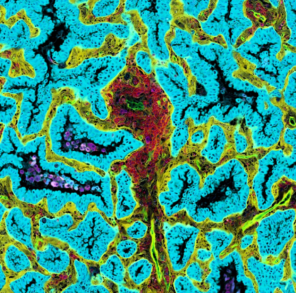
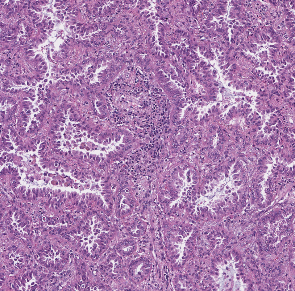 Input: H&E
Output: Virtual IF
Input: H&E
Output: Virtual IF
- CK
- CD45
- CD31
- CD68
- SMA
Virtual multiplex immunofluorescence (IF), lung
Drag to compare the H&E whole slide image input with the virtual multiplex IF.
316
NSCLC whole slide images
150M+
Cell-level annotations
5
Virtual biomarker models
0.90–0.91
ROC AUC
applications
Biomarker Discovery
Identify and validate new biomarker candidates from tissue you’ve already collected.
ADC Target Assessment
Quantify target expression and co-expression patterns to support target selection.
Immuno-Oncology Research
Characterize tumor-immune interactions across a multiplex panel, identifying spatial correlates of response or resistance.
Retrospective Trial Analysis
Apply the panel to archival cohorts without requiring additional physical stains.
Resources
Read More
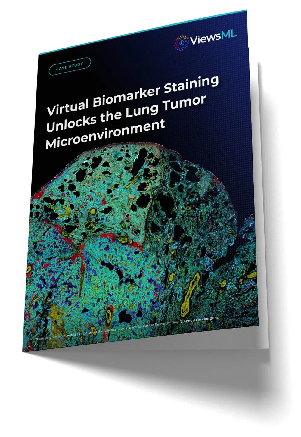
Send us one H&E whole slide image. We return the virtual IHC alongside it.
See virtual staining on your own tissue.
GET STARTED
Tell us
The marker and tissue you are working on.
Send
One H&E whole slide image.
Review
Walk through the virtual IHC with our team.