
pathology WORKFLOW
Get more from the H&E you already have.
A biopsy has to serve diagnosis, subtyping, and downstream molecular testing, and every physical immunohistochemistry (IHC) stain ordered consumes another section of that same finite tissue. The stains ordered need to be balanced with the need to conserve tissue for the molecular testing that comes next.
Virtual staining predicts biomarker expression from the digitized H&E slide already on file, before a physical stain is ordered.
For Research Use Only.
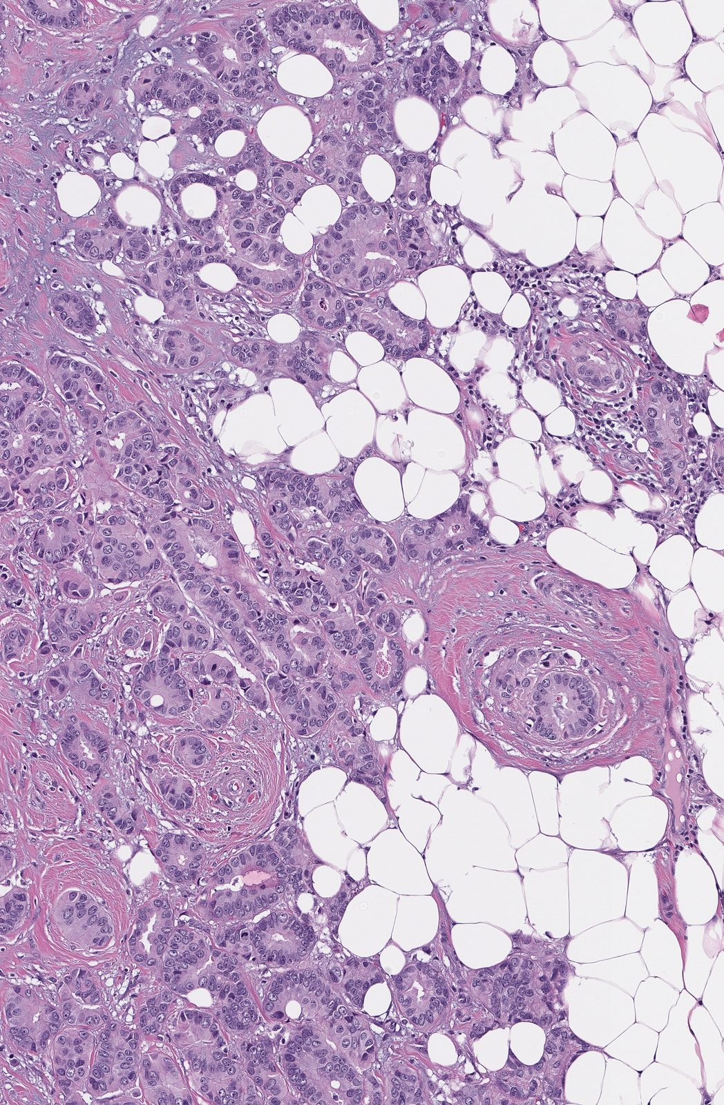
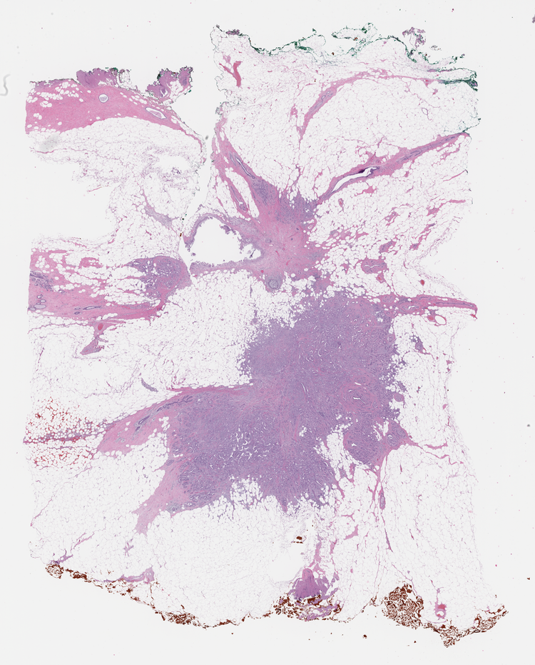
Input
H&E whole slide image.
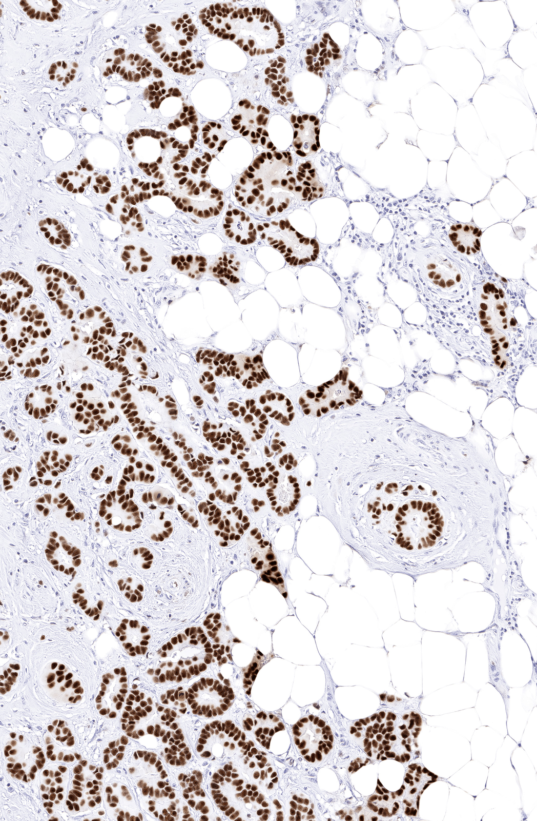
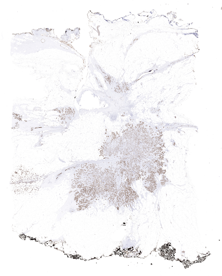
Output
Virtual IHC, generated computationally.
Reference standard
Physical IHC, used as the reference for scoring.
Model performance
0.92
ROC AUC, per cell, across an evaluation set
29.5%
Cell positivity, virtual IHC
148,447
Cells detected
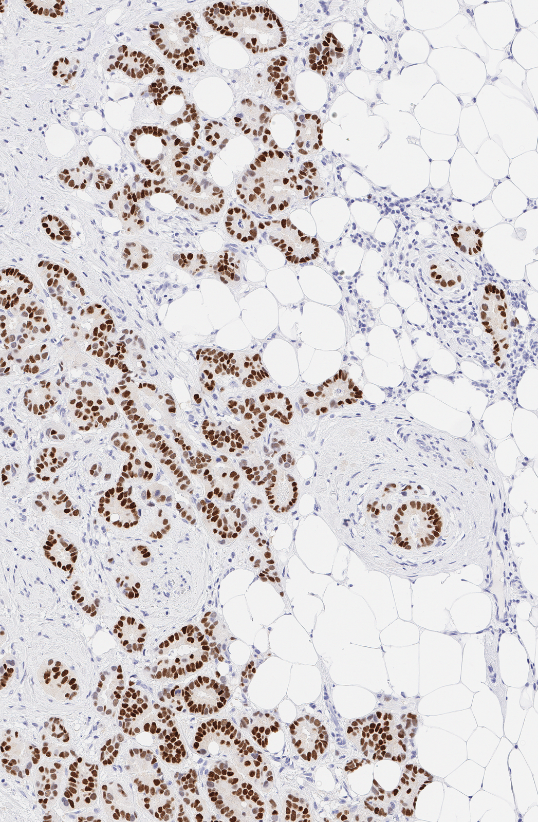
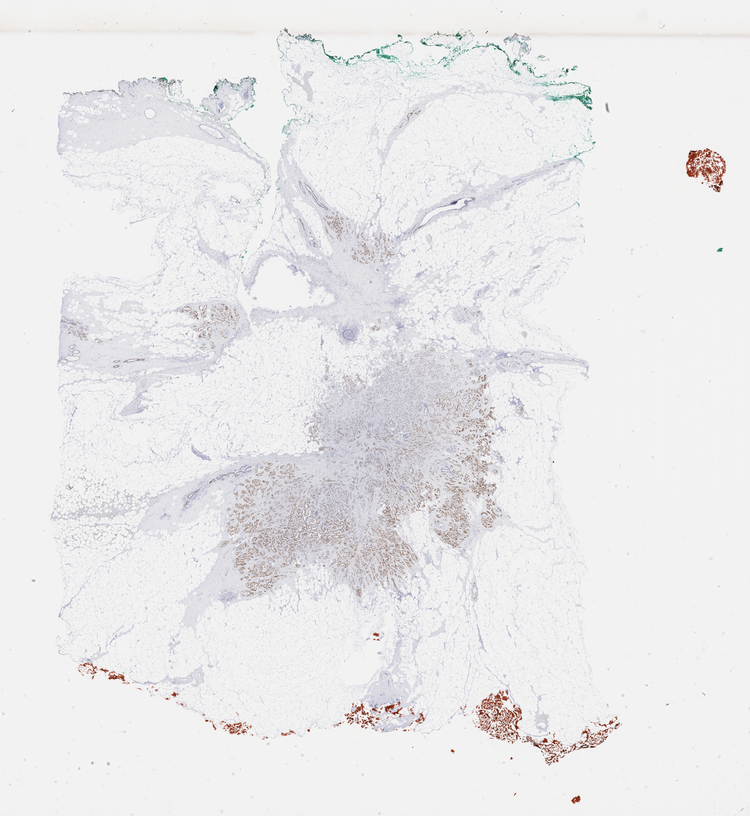
Estrogen Receptor (ER), breast carcinoma · measured across the whole slide
Virtual IHC generated for estrogen receptor (ER) and progesterone receptor (PR) in breast tissue showed high concordance with matched physical IHC, with accurate per-cell localization of biomarker expression.
Virtual ER and PR staining on breast tissue, delivers biomarker status without spending tissue.
THE EVIDENCE
This approach makes it possible to reduce turnaround time and support earlier treatment decisions.
ADVANTAGES
Preserve Tissue
Use virtual staining to guide a more targeted physical IHC panel, instead of staining speculatively.
Reduce Turnaround Time
Get subtyping-relevant information in minutes, from the H&E you already have.
Reduce QNS Risk
Keep tissue that would have been consumed in IHC staining for downstream molecular testing.
Keep Your Current Workflow
Use the digitized H&E already generated in your workflow, no additional steps required.
Resources
Read More
See virtual staining on your own tissue.
GET STARTED
Send us one H&E whole slide image. We return the virtual IHC alongside it.
Tell us
The marker and tissue you are working on.
Send
One H&E whole slide image.
Review
Walk through the virtual IHC with our team.