
Characterize your archive for biomarker expression profiles without consuming tissue.
TISSUE BANKING
Identifying tissue blocks with the desired biomarker profile traditionally means cutting and staining sections, consuming valuable tissue even from blocks that ultimately don't have the requested profile.
Virtual staining generates biomarker expression data directly from the H&E already on file, so archives can be searched and characterized before a single block is cut.
For Research Use Only.
Virtual ER and PR staining matched physical IHC, at the cell level.
THE EVIDENCE
Virtual IHC generated for estrogen receptor (ER) and progesterone receptor (PR) in breast tissue showed high concordance with matched physical IHC, with accurate cell-level localization of biomarker expression.
This approach makes it possible to characterize existing H&E for ER and PR, and select only cases that match the requested profile for physical staining.


Input
H&E whole slide image.


Output
Virtual IHC, generated computationally.
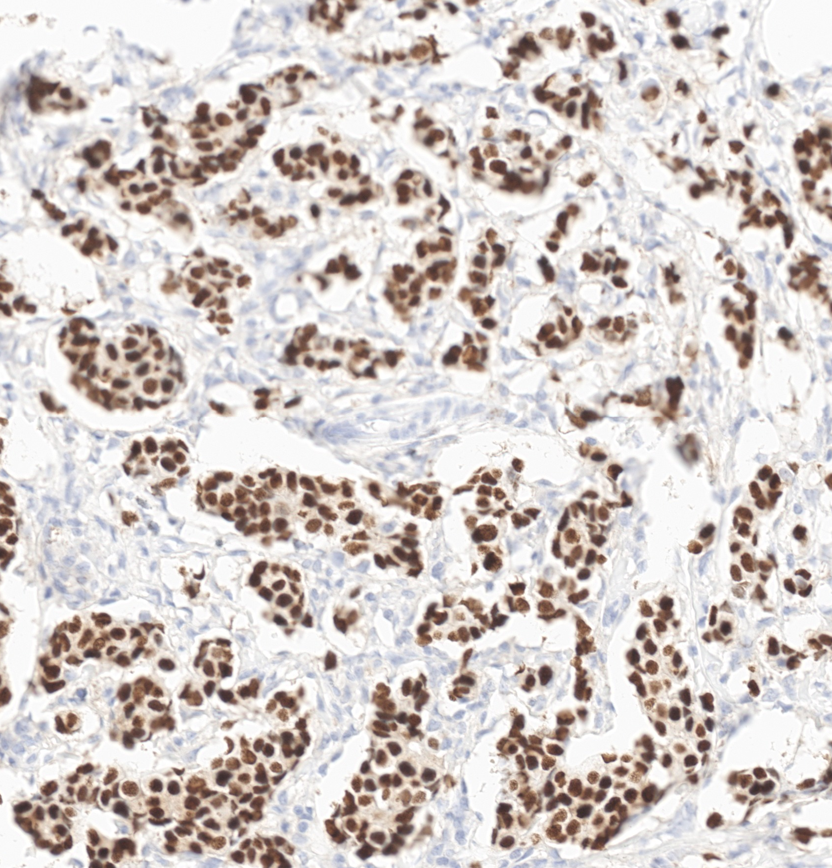

Reference standard
Physical IHC, used as the reference for scoring.
Model performance
0.92
ROC AUC, per cell, across an evaluation set
17.5%
Cell positivity, virtual IHC
24,673
Cells detected
Progesterone Receptor (PR), breast carcinoma · measured across the whole slide
ADVANTAGES OF VIRTUAL STAINING
Preserve Tissue
Search and characterize tissue directly from the digitized H&E you already have, without cutting blocks to find out if they qualify.
Reduce Turnaround Time
Respond to marker-specific requests faster and more precisely.
Increase Inventory Value
Turn an uncharacterized archive into searchable, biomarker-annotated inventory.
Resources
Read More
Send us one H&E whole slide image. We return the virtual IHC alongside it.
See virtual staining on your own tissue.
GET STARTED
Tell us
The marker and tissue you are working on.
Send
One H&E whole slide image.
Review
Walk through the virtual IHC with our team.